A major new study has found that radiation from medical imaging, such as CT scans and X-rays, is associated with a small but measurable increase in the risk of blood cancers in children and adolescents.
The research, published in the New England Journal of Medicine and funded by the National Cancer Institute and National Institutes of Health, analyzed the medical records of nearly 4 million young patients across the U.S. and Canada.
Researchers concluded that about 10% of pediatric blood and bone marrow cancers, roughly 3,000 cases, could be linked to radiation exposure from medical imaging. The risk rose in proportion to the total amount of radiation children received over time.
“Children are particularly vulnerable to radiation-induced cancer due to their heightened radiosensitivity and longer life expectancy,” said Dr. Rebecca Smith-Bindman, lead author of the study and professor of epidemiology and radiology at the University of California, San Francisco. She added that while imaging is often lifesaving, the findings show the need to carefully evaluate and minimize radiation exposure to protect children’s long-term health.
The investigation is the first large-scale North American study to directly measure leukemia and lymphoma risks linked to medical imaging.
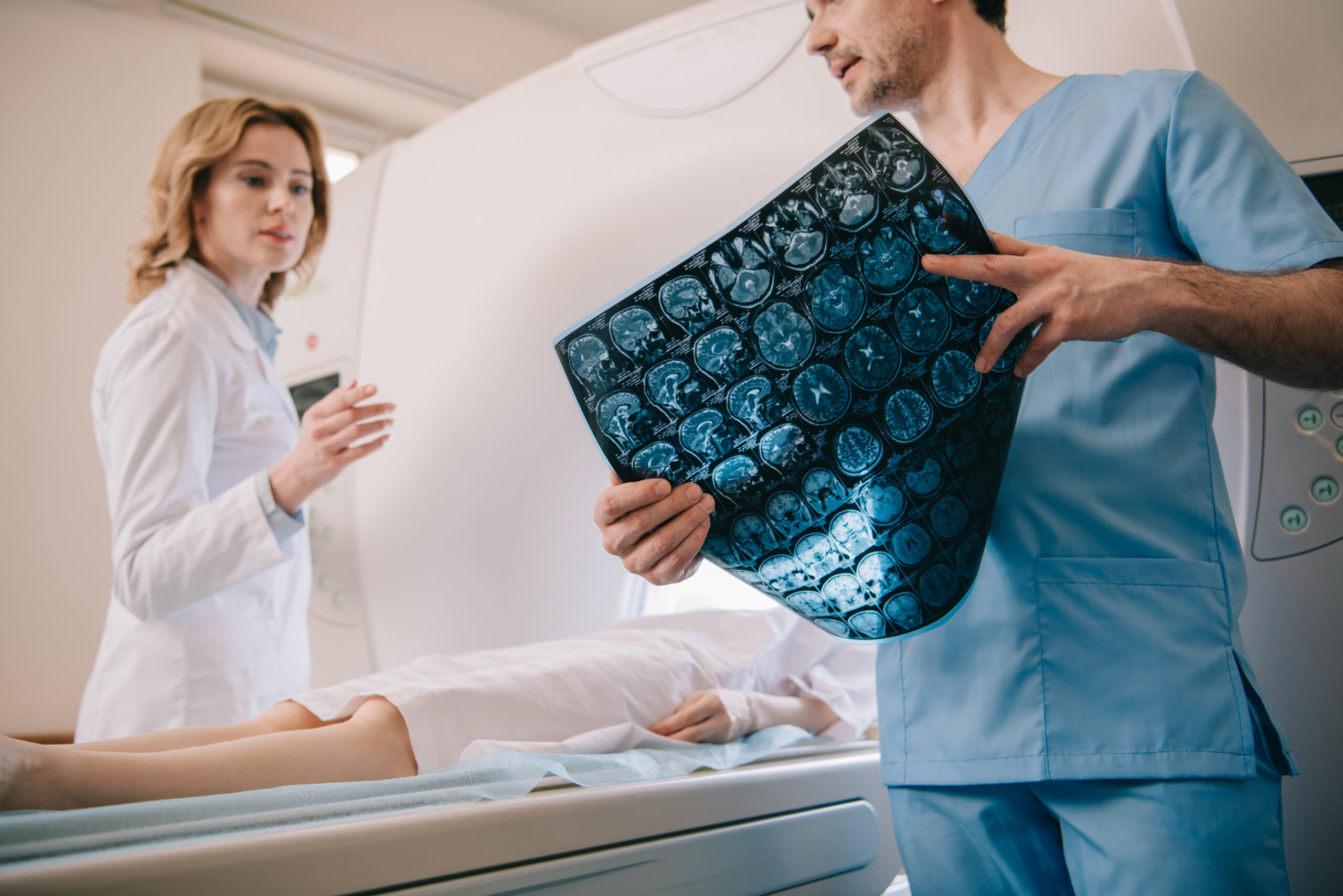
Researchers tracked the imaging histories of 3.7 million children treated between 1996 and 2016. They found a clear dose-response relationship: children who received one or two head CT scans were 1.8 times more likely to develop a hematologic cancer, while those who had multiple scans faced a 3.5-fold increase in risk.
Although CT scans account for a greater share of total radiation exposure, other imaging methods, including radiography, nuclear medicine, and fluoroscopy, were also factored into the study’s bone marrow dose calculations.
The highest doses were associated with head and neck CT scans, where average exposure was about 30.8 milligray. The incidence of blood cancers by age 21 was 0.3% among children with doses above that level, but fewer than 1% of patients in the study reached that amount.
Wesley Bolch, a professor of biomedical and radiological engineering at the University of Florida and a co-author of the study, explained that his team used 3D anatomical models to reconstruct radiation doses for each patient based on their size, sex, and imaging details. Previous cancer risk models often relied on data from atomic bomb survivors, which differ greatly from diagnostic radiation exposure.
“This is the very first study of its kind in the U.S. and Canada, and the very first where each patient was considered in a unique manner,” Bolch said.
The study also found that the risks varied by imaging type. CT scans, which deliver higher doses, were linked to more cancers than radiographs, which use lower levels of radiation. Chest X-rays were the most common imaging test performed, while head CT scans were the most frequent type of CT.
Researchers stressed that medical imaging remains a vital tool for diagnosing disease and guiding treatment. They emphasized that the benefits of imaging far outweigh the risks when scans are medically justified and performed using the lowest possible radiation dose.
“Everybody went into action to figure out what is the appropriate lowest dose of radiation that would give us a good image,” Bolch said. “These risks are low, and when justified by the imaging physician, patient benefits—such as disease detection—will greatly outweigh these very small risks.”
The authors said that reducing unnecessary scans, optimizing radiation doses, and considering alternatives like ultrasound or MRI could help prevent thousands of cancer cases in the future. “Our findings align with international research showing that children are especially radiosensitive,” said Dr. Diana Miglioretti, chief of biostatistics at UC Davis Health. “It’s crucial for clinicians to weigh the immediate benefits of imaging against potential long-term risks.”
The study’s conclusion is clear: medical imaging is essential, but careful use and ongoing improvements in technology are key to minimizing risks for young patients.